Explore facial anatomy for aesthetic medicine, including essential muscles, nerves, vessels, and fat compartments for safer aesthetic treatment planning.
Facial anatomy for aesthetic medicine is the foundation of informed assessment, responsible treatment planning, and a deeper understanding of facial appearance. Beneath the skin lies an interconnected arrangement of muscles, fat compartments, connective tissues, nerves, blood vessels, and bone. These structures work together to support expression, facial movement, and individual features.
For clinicians studying aesthetic procedures, understanding the face as a layered, dynamic structure is more valuable than memorizing isolated landmarks. Facial muscles influence expression lines, soft tissues contribute to contour, and vascular structures create important safety considerations. Anatomical variation also means that a feature appearing similar in two patients may have different underlying relationships.
This guide introduces essential facial anatomy for aesthetic medicine, focusing on structural layers, facial expression muscles, and the relationships between nerves and blood vessels. It provides educational background rather than procedural injection instructions. Clinical treatment requires appropriate professional training, individual assessment, and knowledge of potential complications.
Why Facial Anatomy for Aesthetic Medicine Matters
Modern aesthetic medicine involves more than changing visible lines or contours. It requires understanding how tissues are arranged, how facial expressions are produced, and how age-related changes affect different regions. The skin is only the most visible component of a complex anatomical system.
A sound anatomical foundation helps practitioners interpret facial movement, distinguish static features from dynamic changes, and recognize why treatment plans should be individualized. It also supports informed discussions about limitations, realistic outcomes, and potential adverse effects.
Understanding the Face as a Dynamic Structure
Facial anatomy for aesthetic medicine should be understood as a dynamic system rather than a collection of static anatomical structures. Muscles contract during smiling, speaking, blinking, and other everyday activities. Their actions affect the overlying skin and interact with adjacent tissues. Facial expression muscles are largely supplied by the facial nerve, while muscles of mastication receive motor innervation primarily through the mandibular division of the trigeminal nerve.
These distinctions matter because facial movement is produced by coordinated muscle groups rather than single isolated structures. The appearance of a smile, for example, reflects activity around the mouth and cheeks together with underlying support and soft-tissue distribution.
Anatomical Variation and Individual Assessment
Although textbooks describe typical structures and pathways, facial anatomy for aesthetic medicine requires an understanding of the natural anatomical variations found between individuals. Differences in bone shape, tissue thickness, muscle relationships, and vascular branching contribute to individual appearance. Age, previous procedures, and medical history may further influence the anatomy encountered during assessment.
For this reason, a diagram should be treated as a learning resource rather than a precise map of every patient’s anatomy. The ability to recognize uncertainty and anatomical variation is an important part of responsible aesthetic practice.
Understanding the Main Layers and Structures of the Face
One of the most useful ways to study facial anatomy for aesthetic medicine is to consider the face in layers. The precise arrangement differs between regions, but the skin, subcutaneous tissue, muscular and fascial structures, deeper fat, retaining structures, and facial skeleton all contribute to appearance and function.
Skin and Superficial Soft Tissue
The skin includes the epidermis and dermis, which provide protection, sensation, and structural support. Skin thickness and elasticity differ across facial regions. The delicate periorbital area, for example, has different characteristics from the thicker tissues found in portions of the cheek and lower face.
Immediately beneath the skin, superficial fatty tissue contributes to facial fullness and surface contour. Changes in skin quality and soft-tissue support may alter how underlying muscle activity becomes visible. This relationship helps explain why similar facial movements can produce different patterns of visible lines.
Facial Fat Compartments and Contour
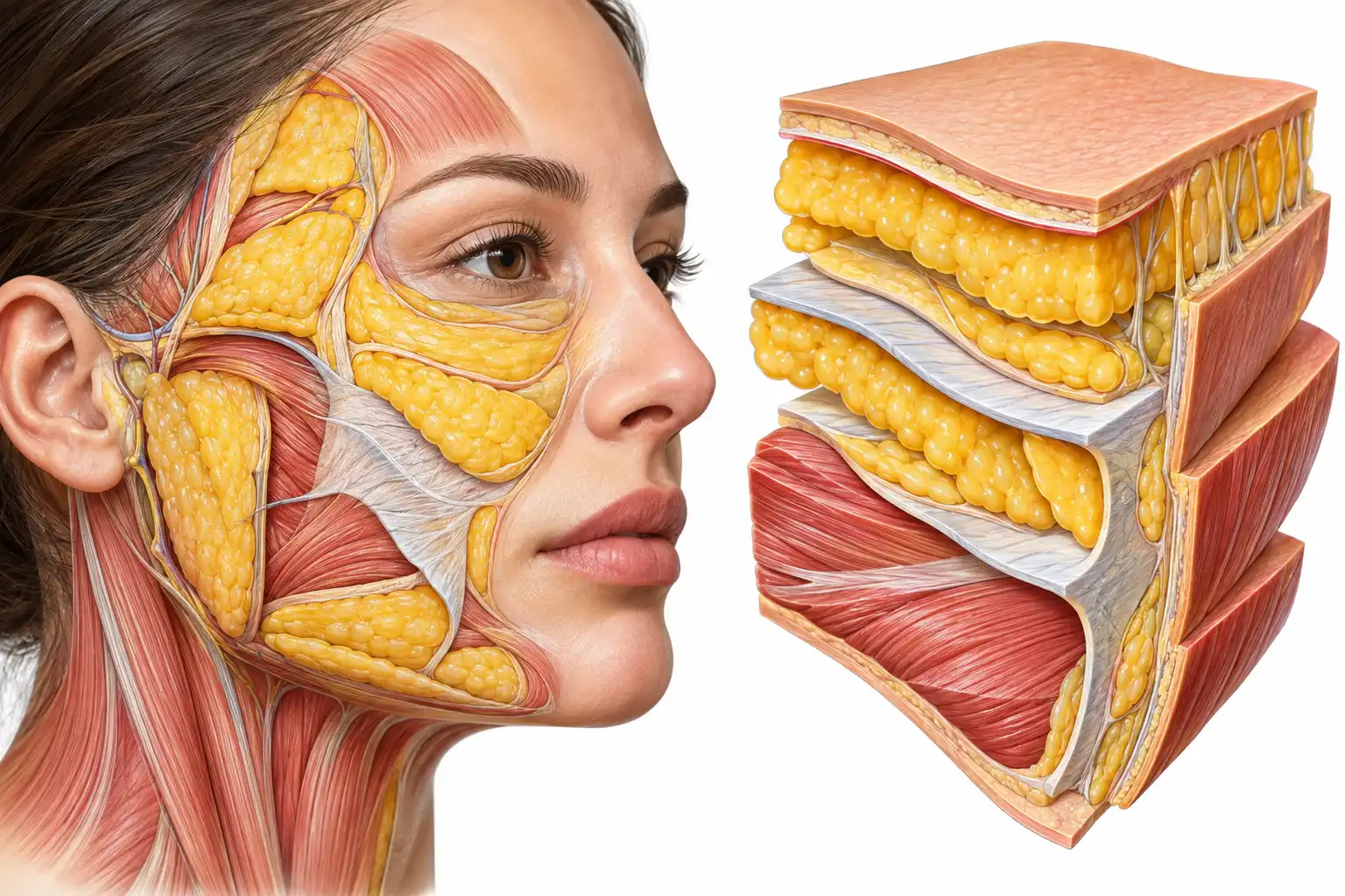
In facial anatomy for aesthetic medicine, facial fat is understood as a collection of distinct compartments rather than a single uniform layer. Superficial and deep fat compartments occupy different anatomical spaces and contribute to the three-dimensional shape of the face. Their distribution influences the cheeks, temples, midface, and lower facial contour.
With aging, facial appearance can change through a combination of soft-tissue redistribution, changes in fat volume, skin alterations, and skeletal remodeling. These processes do not occur identically in every person. Understanding facial fat compartments therefore supports a more complete assessment of facial proportions.
Connective Tissue, Retaining Ligaments, and the SMAS
Connective tissues organize and support facial structures. Retaining ligaments help anchor soft tissues, while the superficial musculoaponeurotic system, commonly called the SMAS, forms an important fibromuscular layer in parts of the face. Its relationships with muscles, fascia, and adjacent tissues vary by region.
These structures are particularly relevant when studying facial support and the visible effects of aging. They also demonstrate why facial anatomy cannot be accurately represented as a series of perfectly uniform horizontal layers throughout the entire face.
Facial Bones and Structural Support
Understanding the facial skeleton is an important part of facial anatomy for aesthetic medicine because it provides the underlying framework that supports facial soft tissues. The maxilla, mandible, zygomatic bones, nasal bones, and surrounding orbital structures contribute to facial proportions and landmarks. Bone shape influences the projection and relationships of the overlying tissues.
Age-related skeletal changes may contribute to alterations in facial support alongside changes in fat, skin, and connective tissue. Recognizing the interaction between these components is more informative than attributing every visible change to a single tissue layer.
Essential Facial Muscles and Their Functions
Understanding facial muscles is a central component of facial anatomy for aesthetic medicine, particularly when studying expression patterns and facial movement. Most muscles of facial expression are innervated by the facial nerve, or cranial nerve VII. Unlike many skeletal muscles, several facial expression muscles attach to skin or adjacent soft tissues, allowing contraction to produce visible movement.
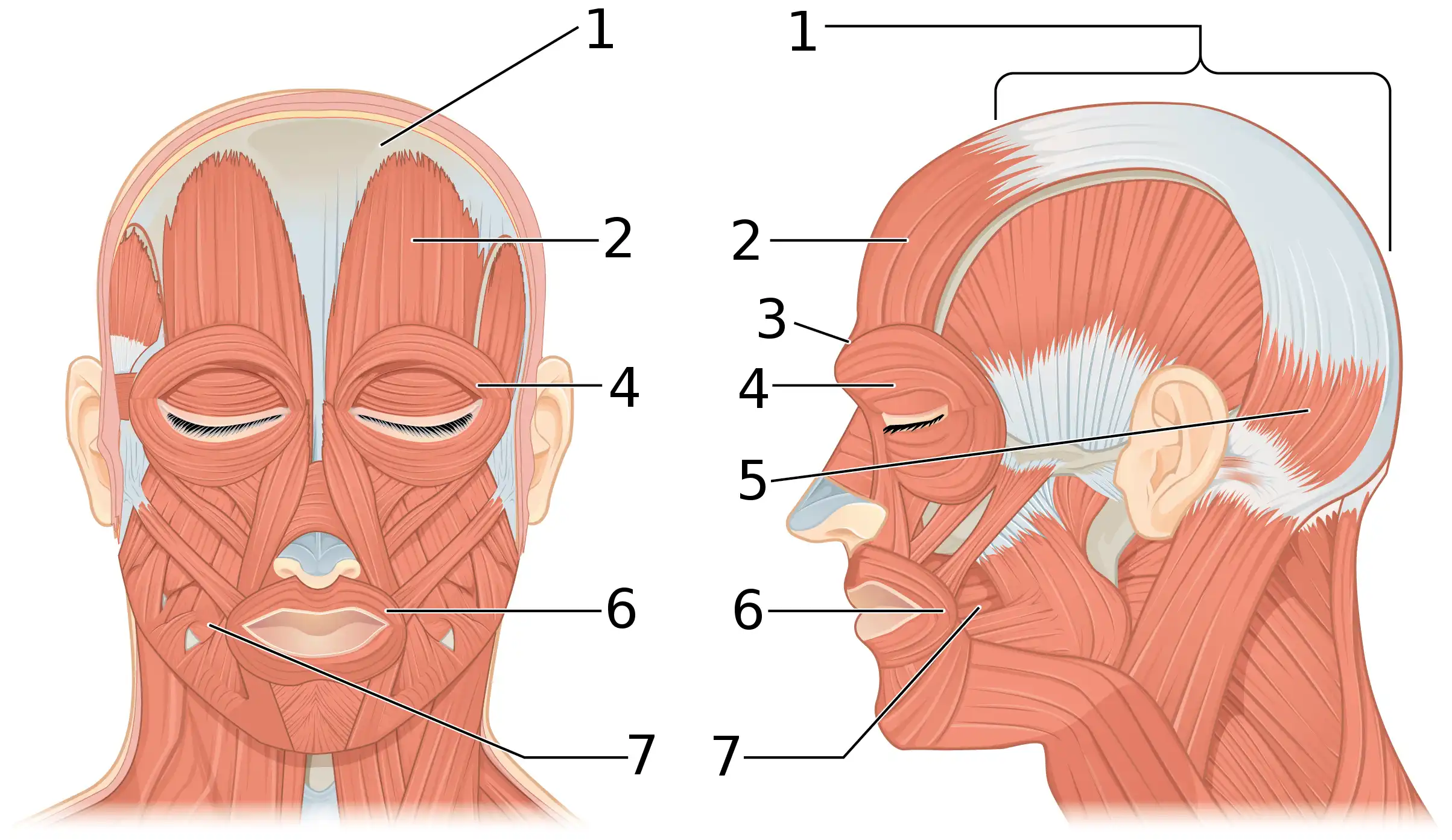
These muscles operate in coordinated groups. Their position, direction of pull, and interaction with surrounding tissues influence expression patterns. The following overview focuses on commonly studied muscles and their general functions rather than procedural treatment locations.
Frontalis and the Forehead
The frontalis is the frontal belly of the occipitofrontalis muscle. It elevates the eyebrows and contributes to horizontal forehead lines during expression. Its activity interacts with muscles that lower or draw the eyebrows inward, creating a balance of movement across the upper face.
Forehead appearance therefore depends on more than the strength of the frontalis alone. Eyebrow position, skin characteristics, and the activity of adjacent muscles all influence visible movement.
Corrugator Supercilii and Procerus
The corrugator supercilii draws the eyebrows medially and downward, contributing to vertical glabellar lines. The procerus acts in the region between the eyebrows and over the upper nose, contributing to transverse skin wrinkling near the nasal root.
These muscles are often discussed together because their combined activity affects expression in the glabellar region. Understanding their functions provides a basis for interpreting dynamic facial lines without assuming that every visible crease has the same anatomical cause.
Orbicularis Oculi and Eye Closure
The orbicularis oculi surrounds the eye and plays a central role in eyelid closure. Its palpebral portion supports gentle blinking, while the orbital portion contributes to stronger closure. Muscle activity around the lateral eye can also produce expression-related skin folds.
The periorbital region contains closely related muscles, nerves, blood vessels, and delicate soft tissues. Its complex anatomy illustrates why detailed regional knowledge is essential in aesthetic education.
Zygomaticus Major and Minor
The zygomaticus major helps elevate the corner of the mouth during smiling, while the zygomaticus minor assists in elevating the upper lip. Their actions work alongside other muscles of the cheek and mouth to create natural facial expressions.
Differences in muscle activity and soft-tissue anatomy contribute to variations in smile shape. Aesthetic assessment should therefore consider movement and facial symmetry in addition to appearance at rest.
Orbicularis Oris and Perioral Movement
The orbicularis oris is a complex muscular structure surrounding the mouth. It contributes to lip closure, pursing, and movements involved in speech and facial expression. Its function depends on coordination with neighboring muscles that elevate, depress, or retract the lips and oral commissures.
The mouth is a highly mobile region, and its appearance changes continuously during speech and expression. Understanding this functional complexity helps explain why perioral anatomy should be studied as an interconnected system.
Masseter, Mentalis, and Lower Facial Function
The masseter is a major muscle of mastication that elevates the mandible. Unlike the muscles of facial expression, it receives motor innervation from the mandibular division of the trigeminal nerve. Its size and activity can influence the appearance of the lower facial contour.
The mentalis contributes to elevation and protrusion of the lower lip and wrinkling of the chin skin. Together with the muscles surrounding the mouth, it participates in expressions involving the lower face.
Studying these muscles alongside the facial skeleton, connective tissues, and vascular anatomy helps build a more complete understanding of lower facial structure.
For additional anatomical reference, consult the NCBI Bookshelf overview of facial muscles and the NCBI Bookshelf review of facial artery anatomy. These resources provide further detail on muscle function, innervation, vascular relationships, and clinical relevance.
5. What are facial fat compartments, and how do they affect aging?
1. Why is facial anatomy important in aesthetic medicine?
Facial anatomy for aesthetic medicine helps healthcare professionals understand the relationships between muscles, nerves, blood vessels, fat compartments, and underlying bones. This knowledge supports comprehensive facial assessment, individualized treatment planning, and recognition of potential anatomical risks. Because facial structures vary between individuals, anatomical education must be combined with appropriate clinical training and patient-specific evaluation.
2. Which facial muscles are most important in aesthetic procedures?
Important facial muscles include the frontalis, corrugator supercilii, procerus, orbicularis oculi, orbicularis oris, zygomaticus major, mentalis, and masseter. These muscles influence eyebrow movement, smiling, eye closure, lip movement, and lower facial contour. Understanding facial muscles anatomy helps clinicians distinguish expression-related changes from other structural characteristics of the face.
3. What are the main anatomical layers of the face?
The face contains skin, subcutaneous tissues, superficial and deep fat compartments, muscles, fascia, connective tissues, and underlying bone. Their arrangement differs across anatomical regions. The superficial musculoaponeurotic system (SMAS) and retaining ligaments also contribute to facial support. Understanding these relationships is essential when studying facial anatomy for aesthetic procedures.
4. Why is facial vascular anatomy important for dermal filler treatments?
Facial vascular anatomy is important because facial arteries and veins have variable pathways and relationships with surrounding tissues. Accidental injection into a blood vessel can cause serious complications, including tissue ischemia and, in rare cases, vision loss. Knowledge of vascular anatomy is therefore a fundamental safety requirement, but anatomical knowledge alone cannot eliminate procedural risks.
5. What are facial fat compartments, and how do they affect aging?
Facial fat compartments are distinct regions of superficial and deep adipose tissue that contribute to facial volume, contour, and support. Age-related changes in these compartments may occur alongside alterations in skin elasticity, connective tissues, and bone structure. These combined processes can influence cheek fullness, facial folds, and the appearance of the midface.
6. How does facial anatomy help with aesthetic treatment planning?
Understanding facial anatomy helps qualified practitioners assess facial proportions, muscle activity, tissue characteristics, and anatomical variation. It also supports discussion of treatment limitations and possible complications. Effective planning requires more than identifying visible concerns; it involves medical history, individual assessment, clinical judgment, and appropriate professional training.
7. What is the difference between facial nerves and facial muscles?
Facial muscles generate movement through contraction, while nerves transmit signals that control movement or carry sensory information. The facial nerve primarily supplies the muscles of facial expression. The trigeminal nerve provides most facial sensation and supplies motor fibers to the muscles of mastication through its mandibular division. Understanding these different functions is important in facial anatomy education.
8. Can facial anatomy be learned through online courses alone?
Online courses, anatomical illustrations, and educational articles can provide a valuable theoretical foundation. However, they do not replace supervised practical education, appropriate clinical qualifications, or competency assessment. Healthcare professionals involved in aesthetic procedures should pursue structured training that includes relevant anatomy, complication recognition, patient assessment, and clinical safety principles.
Facial Vascular Anatomy and Its Importance in Aesthetic Medicine
Understanding facial vascular anatomy is a fundamental part of facial anatomy for aesthetic medicine. Blood vessels deliver oxygen and nutrients to facial tissues, support normal healing, and maintain the health of the skin and underlying structures. Their pathways also create important clinical considerations for practitioners involved in minimally invasive aesthetic procedures.
The facial vascular network is complex, interconnected, and anatomically variable. Major arteries may follow recognizable general courses, but their branching patterns and relationships with surrounding tissues can differ significantly between individuals. Consequently, an anatomical illustration cannot reliably predict the precise location of every vessel in a particular patient.
The Facial Artery and Its Branches
The facial artery is a major branch of the external carotid artery. It supplies numerous structures of the face and follows a curved pathway from the lower facial region toward the mouth, nose, and medial eye area. Its branches include vessels supplying the lips and surrounding tissues, while its terminal anatomy can vary.
One important characteristic of the facial artery is its relationship with facial muscles and connective tissues. The vessel may change its relative position as it travels through different regions. This variability makes comprehensive anatomical education particularly important for clinicians studying aesthetic procedures.
For further scientific reading, the NCBI Bookshelf review of facial artery anatomy provides an overview of the vessel’s course, branches, anatomical relationships, and clinical significance.
Superficial Temporal and Infraorbital Vascular Structures
The superficial temporal artery contributes to the blood supply of the temporal and scalp regions. Other vessels, including the infraorbital artery and branches associated with the orbital region, participate in supplying the central and upper face.
These vascular systems form connections with neighboring arterial networks. Their anatomical relationships are relevant when considering facial circulation and understanding how vascular complications may affect tissues beyond the immediate area of a procedure.
Why Vascular Knowledge Matters for Dermal Fillers
Facial anatomy for dermal fillers requires an understanding of vascular pathways, anatomical variation, and potential complications. Accidental placement of filler material within a blood vessel can obstruct circulation and produce tissue ischemia. In rare but serious circumstances, vascular complications may involve vision loss or stroke.
The U.S. Food and Drug Administration’s dermal filler safety guidance explains these risks and emphasizes appropriate professional training, anatomical knowledge, patient assessment, and complication preparedness.
Importantly, knowledge of a typical vascular pathway does not guarantee procedural safety. Clinicians must recognize that individual anatomical differences, previous treatments, and other patient-specific factors may influence risk.
Facial Nerve Anatomy and Functional Relationships
Understanding facial nerves is an essential part of facial anatomy for aesthetic medicine because these structures support sensation, movement, and normal facial function. Understanding their distribution helps healthcare professionals distinguish motor activity from sensory pathways and appreciate the consequences of injury to different anatomical structures.
Two cranial nerves are especially important in introductory facial anatomy: the facial nerve and the trigeminal nerve. Although both are associated with the face, their principal functions differ.
The Facial Nerve: Motor Control of Expression
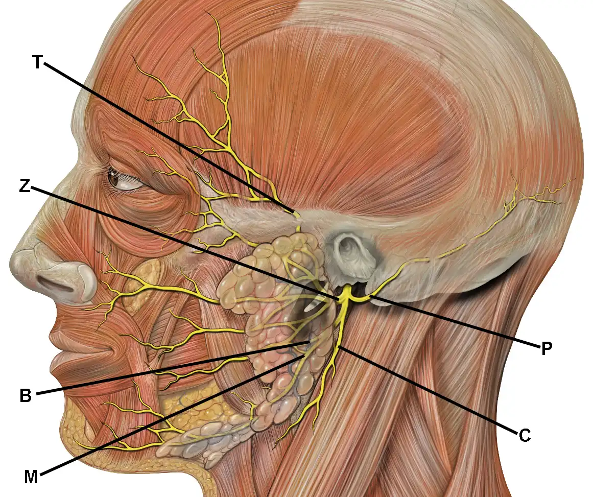
The facial nerve, also known as cranial nerve VII, supplies most muscles responsible for facial expression. Its motor branches are commonly described as temporal, zygomatic, buccal, marginal mandibular, and cervical branches.
These branches contribute to movements such as eyebrow elevation, eye closure, smiling, and lower lip activity. Because facial expressions depend on coordinated muscle activation, impairment of motor function can affect facial symmetry and everyday activities.
The relationships between facial nerve branches and surrounding structures vary across regions. Understanding these relationships is relevant to anatomical education, surgical assessment, and recognition of possible nerve-related complications.
The Trigeminal Nerve: Facial Sensation and Mastication
The trigeminal nerve, or cranial nerve V, provides most somatic sensation to the face. Its three major divisions are the ophthalmic, maxillary, and mandibular nerves.
The ophthalmic division supplies sensory information from areas including the forehead and upper facial region. The maxillary division contributes sensation to the midface, while the mandibular division supplies sensory territories of the lower face and carries motor fibers to the muscles of mastication.
Understanding these divisions helps explain why facial sensation and facial movement should not be treated as identical functions. It also provides useful background for interpreting neurological symptoms and anatomical relationships.
Why Nerve Preservation Matters
In facial anatomy for aesthetic medicine, understanding nerve function is important because facial nerves contribute to eye protection, oral movement, communication, and facial expression.
Sensory pathways are equally important for detecting touch, pain, and other stimuli.
In aesthetic medicine, awareness of nerve anatomy supports more comprehensive assessment and informed discussion of potential complications. Persistent weakness, unexpected sensory changes, or other neurological symptoms following a procedure require appropriate medical evaluation.
Comparing Facial Muscles, Fat Compartments, and Bone Structure
A comprehensive understanding of facial anatomy for aesthetic medicine requires distinguishing between structures that produce movement, provide volume, and support facial shape. Although these components interact continuously, their primary anatomical roles differ.
Facial Muscles: Movement and Dynamic Expression
Facial muscles generate movements associated with smiling, frowning, blinking, and speaking. Repeated contraction can contribute to dynamic expression lines, particularly in areas such as the forehead and around the eyes.
However, visible wrinkles cannot always be explained by muscle activity alone. Skin characteristics, environmental exposure, connective tissue changes, and aging also influence their appearance.
Facial Fat Compartments: Volume and Contour
Superficial and deep facial fat compartments contribute to fullness, transitions between facial regions, and overall contour. Their anatomical organization influences how the face appears both at rest and during movement.
Age-related changes may involve alterations in compartment volume, position, and surrounding support. These changes can occur alongside skin and skeletal remodeling, producing differences in facial appearance that cannot be attributed to fat loss alone.
Facial Bones: Projection and Structural Framework
The facial skeleton provides structural support beneath the soft tissues. The maxilla, mandible, zygomatic bones, and orbital margins contribute to facial proportions and the relationships between different regions.
Skeletal anatomy is particularly important when studying facial projection, symmetry, and changes associated with aging. Soft-tissue appearance must therefore be interpreted in relation to the underlying framework rather than as an isolated surface feature.
How These Structures Work Together
Consider the appearance of the midface. Its contour reflects the interaction of skeletal projection, superficial and deep fat compartments, retaining structures, skin quality, and muscular movement. A visible change in one area may therefore involve several anatomical components.
This layered perspective supports more thoughtful facial assessment. Rather than assuming that one treatment addresses every visible concern, qualified clinicians can consider the underlying anatomy, the patient’s goals, and the limitations of available interventions.
Best Practices for Studying Facial Anatomy in Aesthetic Medicine
Developing a strong foundation in facial anatomy for aesthetic medicine requires a structured approach to learning that connects anatomical knowledge with clinical relevance. Memorizing muscle names is useful, but it is not enough to understand the relationships that influence facial movement, appearance, and clinical safety.
Study Anatomy by Facial Region
Dividing the face into upper, middle, and lower regions can make complex anatomical information easier to organize. Each region contains characteristic muscles, vascular structures, nerves, connective tissues, and skeletal landmarks.
Regional learning should be followed by studying how these structures connect across boundaries. Facial anatomy is continuous, and anatomical systems do not operate independently simply because they are described in separate textbook chapters.
Use Multiple Educational Resources
Medical textbooks, peer-reviewed anatomical studies, three-dimensional models, and properly labeled illustrations provide complementary learning opportunities. Explore our blog for additional educational articles on facial anatomy and aesthetic medicine. Three-dimensional resources may help learners visualize depth and relationships that are difficult to understand from a single flat diagram.
Nevertheless, digital models and illustrations have limitations. They often depict representative anatomy rather than the full range of normal human variation. Learners should compare sources critically and prioritize accurate, current educational material.
Connect Theoretical Knowledge With Supervised Training
For qualified healthcare professionals pursuing aesthetic procedures, anatomical study should form part of a broader training program. Appropriate education includes patient assessment, contraindications, informed consent, complication recognition, and supervised clinical skill development.
Practical competence cannot be established through reading alone. Professional qualifications, regulatory requirements, and clinical supervision remain essential considerations for anyone intending to perform invasive aesthetic treatments.
Recognize Anatomical Variation
Recognizing normal anatomical variation is one of the most important principles of facial anatomy for aesthetic medicine, particularly when evaluating individual differences in facial structures. The location, branching, and relationships of vessels and nerves may differ from standard illustrations. Previous surgery, trauma, or aesthetic treatment can also alter anatomical relationships.
Recognizing these uncertainties encourages more careful assessment and prevents excessive confidence in simplified anatomical maps. It is especially important when studying regions containing complex vascular connections.
Common Mistakes When Learning Facial Anatomy for Aesthetic Procedures
Several misconceptions can limit the usefulness of anatomical education. Identifying these mistakes early helps learners build a more accurate understanding of facial structures and their clinical significance.
Memorizing Landmarks Without Understanding Tissue Relationships
Knowing the approximate location of a muscle or vessel does not necessarily explain its relationship to surrounding tissues. Facial structures exist at different depths, and these relationships can change across regions.
A more effective approach combines anatomical terminology with an understanding of tissue layers, functional movement, and individual variation.
Assuming Every Face Has Identical Anatomy
Standard anatomical diagrams are valuable teaching tools, but they represent generalized patterns. They should not be interpreted as exact representations of every individual.
Differences in facial proportions, vascular branching, muscle relationships, and soft-tissue distribution demonstrate why individualized assessment remains essential.
Confusing Dynamic Wrinkles With Structural Changes
Dynamic wrinkles are associated with facial muscle activity, while other visible changes may be influenced by skin characteristics, soft-tissue support, and skeletal anatomy. These processes frequently overlap.
Interpreting every facial line as a purely muscular problem oversimplifies the anatomy and may lead to unrealistic expectations about aesthetic interventions.
Underestimating Vascular Complications
One of the most serious mistakes in aesthetic education is assuming that minimally invasive procedures are automatically low risk. Dermal fillers can produce complications ranging from temporary swelling and bruising to uncommon but potentially devastating vascular events.
Warning signs such as unusual pain, skin blanching or discoloration, sudden visual changes, and neurological symptoms require immediate attention. Suspected vascular complications must be managed urgently by appropriately trained medical professionals according to established clinical protocols.
Relying on Online Education as a Substitute for Clinical Competence
Online learning can strengthen theoretical understanding, but completing a digital course does not automatically establish the competence or legal authorization required to perform medical procedures.
Professional training should reflect the learner’s existing qualifications, applicable regulations, supervised experience, and ability to recognize and respond to complications.
Understanding the Cost and Value of Facial Anatomy Education
The cost of education in facial anatomy for aesthetic medicine varies according to the course format, level of specialization, teaching resources, and practical learning components. Introductory online courses may focus on theoretical knowledge, while more advanced programs can include instructor-led teaching, anatomical demonstrations, and supervised educational activities.
Price alone is not a reliable indicator of educational quality. A lower-cost course may provide a useful introduction, whereas a more expensive program is not necessarily appropriate for every learner.
What Should a Quality Anatomy Course Include?
When evaluating courses in facial anatomy for aesthetic medicine, healthcare professionals should consider whether the curriculum addresses facial muscles, vascular anatomy, nerve pathways, fat compartments, skeletal relationships, and clinically relevant anatomical variation.
Additional considerations include instructor qualifications, evidence-based learning materials, clearly defined educational objectives, and transparent information about assessment or certification.
Choosing Education Based on Professional Goals
Beginners may benefit from foundational anatomy education before exploring more specialized topics. Experienced healthcare professionals may require advanced instruction relevant to their existing clinical scope and professional responsibilities.
Before enrolling, learners should confirm whether a course provides theoretical education only or includes supervised practical training. Learn more about our educational approach and learning opportunities on our About Us page. Any certificate should be evaluated in relation to professional licensing and local regulatory requirements.
Conclusion: Building a Strong Foundation in Facial Anatomy for Aesthetic Medicine
Facial anatomy for aesthetic medicine provides the scientific foundation for understanding facial expression, structural support, and the anatomical relationships relevant to aesthetic care. Muscles, nerves, blood vessels, fat compartments, connective tissues, and bones contribute different functions while operating as an interconnected system.
A detailed understanding of these structures supports more informed assessment and highlights the importance of anatomical variation. It also reinforces why patient safety, appropriate qualifications, and professional judgment must remain central to aesthetic practice.
For healthcare professionals interested in developing their knowledge, structured anatomical education is an important starting point. Combining reliable scientific resources with suitable supervised training can help build a stronger foundation for responsible professional development.
Continue Your Professional Learning:
For questions about available educational programs and enrollment requirements, contact our team.
Explore educational opportunities in facial anatomy and aesthetic medicine that match your qualifications and learning goals. Choose programs with clear curricula, appropriately qualified educators, and a strong emphasis on anatomical understanding and patient safety.